Abstract
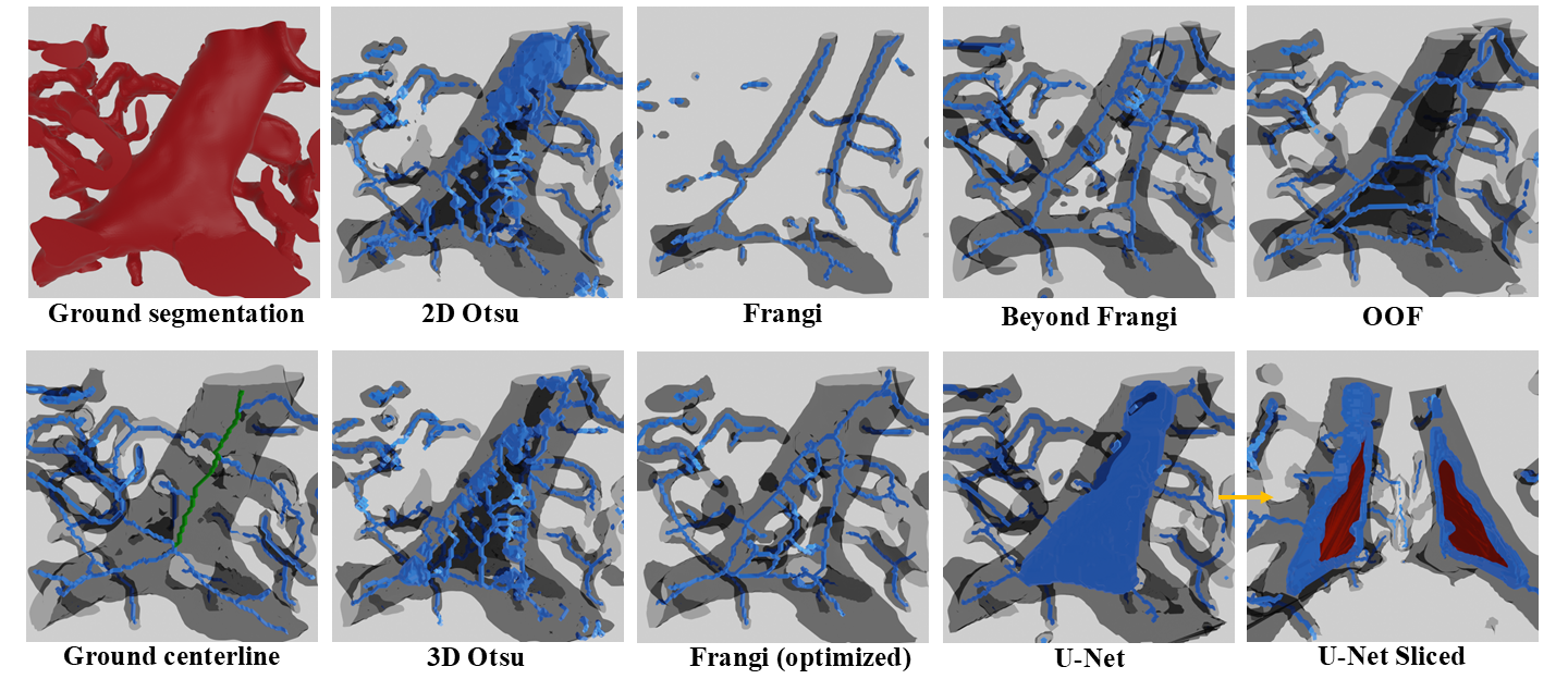
While many algorithms exist for segmenting and skeletonizing vessel-like structures in small-scale images, they are not designed and have not been tested on gigavoxel-scale 3D images. We propose a comprehensive yet compact survey of available algorithms. We focus on essential features for microvascular analysis, including extracting vessel surfaces and the network's associated connectivity. Algorithms were selected based on popularity and availability and provide a thorough quantitative analysis of their performance on data sets acquired using emerging techniques.
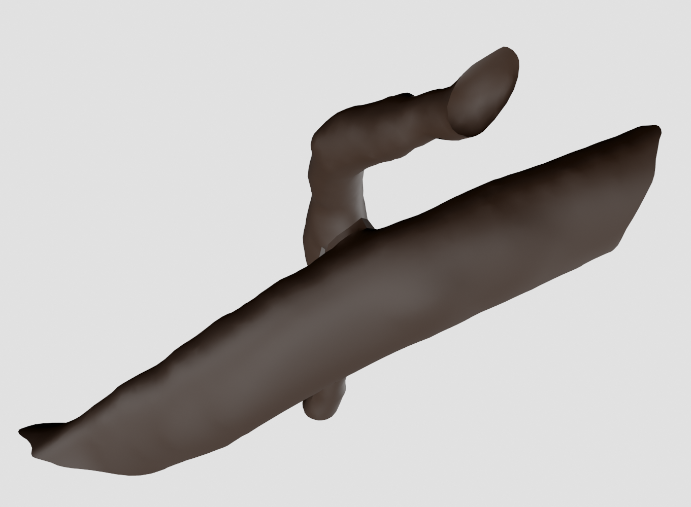
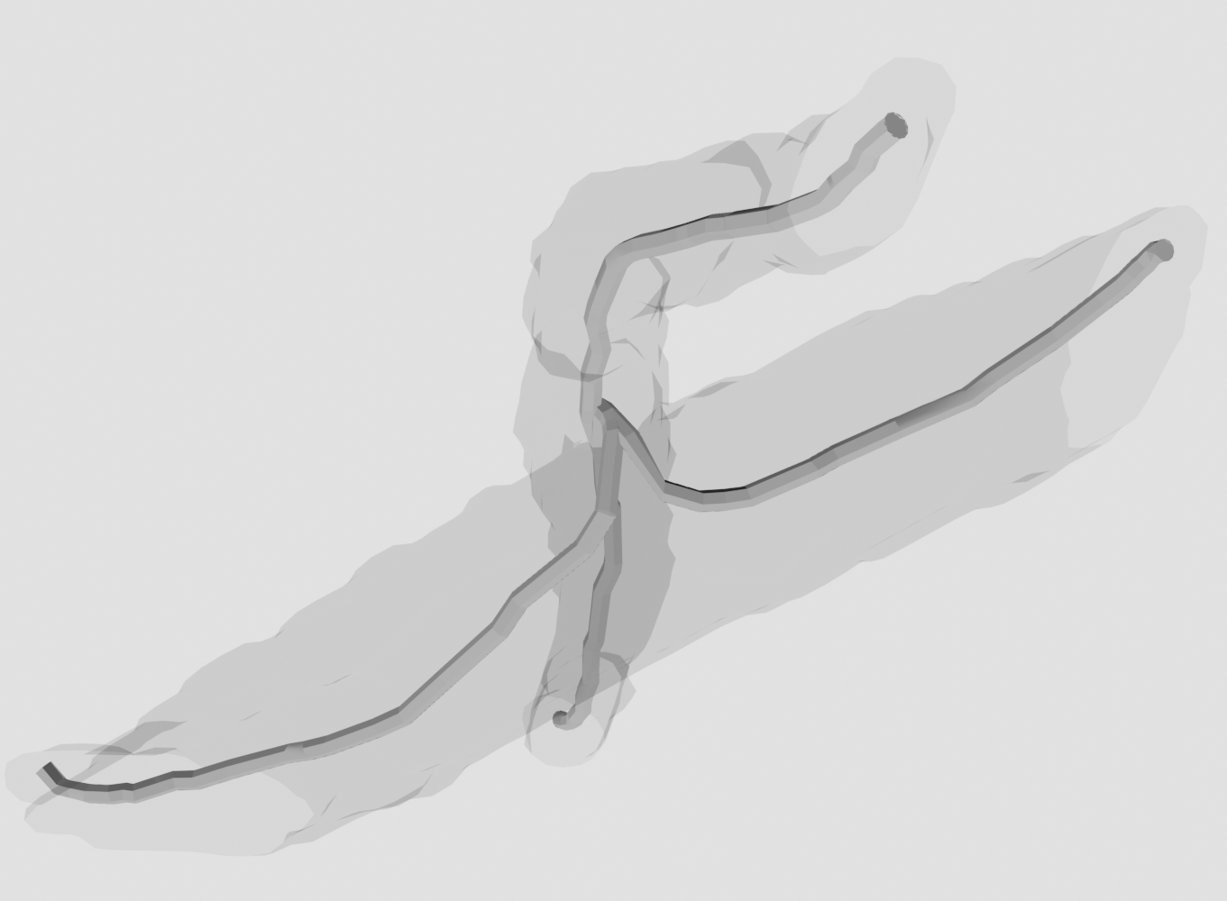
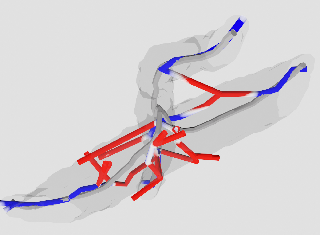
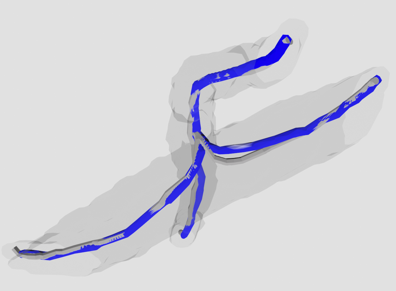
Visualization
- Segmentation
- Skeleton
Resources
Paper: Our paper is available on Frontiers in Bioinformatics and locally.
Code: Our code is available on Github.
Data: Our data (KESM/LSFM/MICRO-CT) is available upon request.
Citation
@article{goharbavang5segmentation,
  title={Segmentation and Modeling of Large-Scale Microvascular Networks: A Survey},
  author={Goharbavang, Helya and Ashitkov, Artem T and Pillai, Athira and Wythe, Joshua D and Chen, Guoning and Mayerich, David},
  journal={Frontiers in Bioinformatics},
  volume={5},
  pages={1645520},
  year={2025},
  publisher={Frontiers}
}